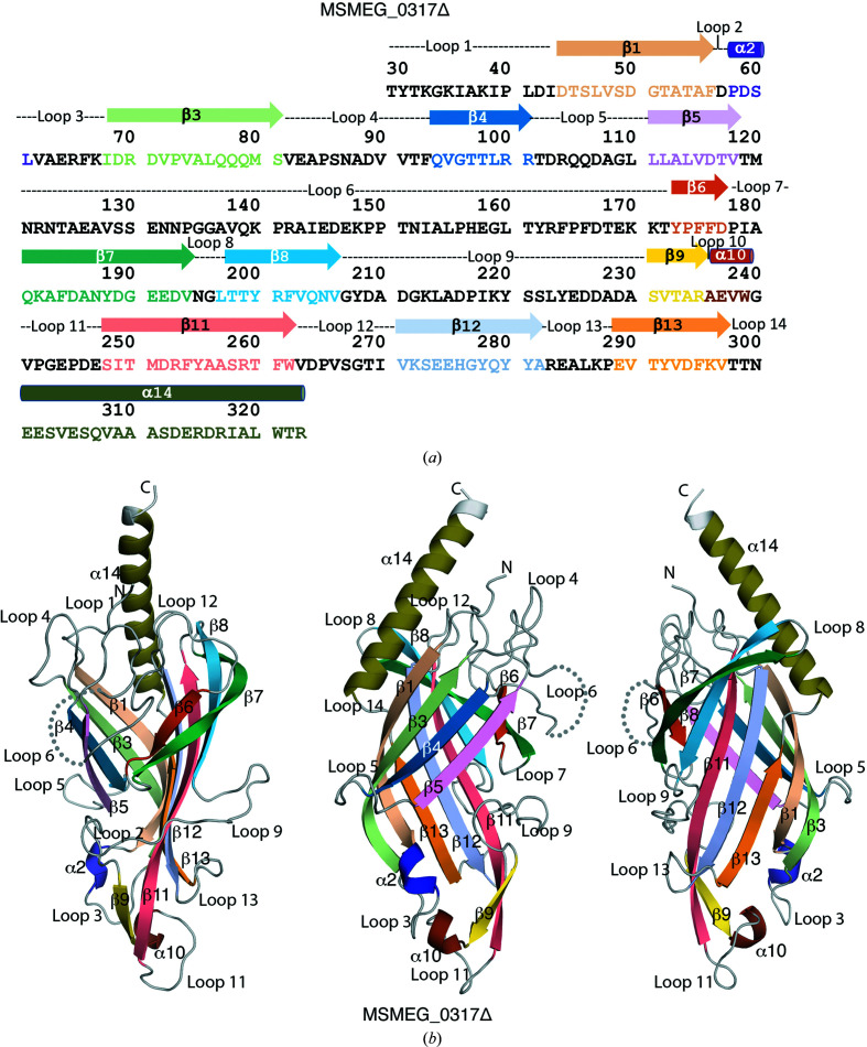
Figure 2.
The amino-acid sequence and the crystal structure of the periplasmic domain of MSMEG_0317Δ. (a) The sequence of MSMEG_0317Δ showing secondary-structure elements derived from the crystal structure of MSMEG_0317Δ. (b) The crystal structure of MSMEG_0317Δ in different views. The secondary-structure elements are colour-coded. The disordered loop 6 is shown by dotted lines. See also Supplementary Figs. S2–S4.

