
Fig. 6. miR-9–5p, miR-203a-3p, and miR-195–5p target the 3’UTR of ONECUT2.
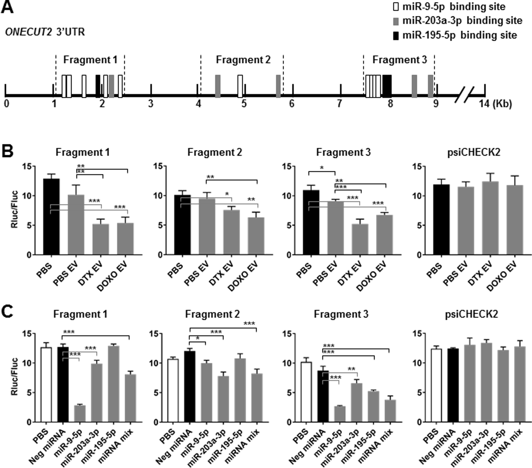
(A) A schematic representative showing putative binding sites of miR-9–5p, miR-203a-3p, and miR-195–5p in the 3’UTR of human ONECUT2 and the regions cloned into the luciferase reporter plasmid constructs. (B) MDA231 cells were transfected with psiCHECK2 reporter plasmids containing indicated ONECUT2 3’UTR region or with the psiCHECK2 vector (2 µg DNA per 2×10⁵ cells). After 12 h, transfected cells were exposed to PBS or EVs from PBS/DTX (4 nM)/DOXO (125 nM)-treated MDA231 cells for 48 h before luciferase activities were measured. Ratio between Renilla luciferase and firefly luciferase activities (Rluc/Fluc) is shown. (C) MDA231 cells were co-transfected with indicated psiCHECK2 reporter plasmids (2 µg DNA per 2×10⁵ cells) and miRNA mimics (individually or with a 1:1:1 mixture of miR-9–5p, miR-203a-3p, and miR-195–5p mimics for a total of 25 pmol). Luciferase activities were analyzed at 48 h. *P<0.05, **P<0.01, ***P<0.001.