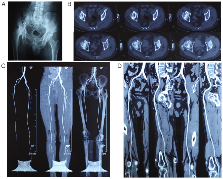
Figure 1.
Imaging results of non-traumatic femoral head necrosis. (A) Representative X-ray showing lack of any joint space in the right hip joint and collapse of the acetabulum and femoral head, along with altered structure. (B) Representative computerized tomography image showing stenosis of the hip joint space, hollow femoral head and abnormal calcification. (C) Angiography of the lower extremities showing insufficient supply from blood vessels in the right femoral head. (D) Angiography of one side of the lower extremity showing properly functioning main blood vessels.

