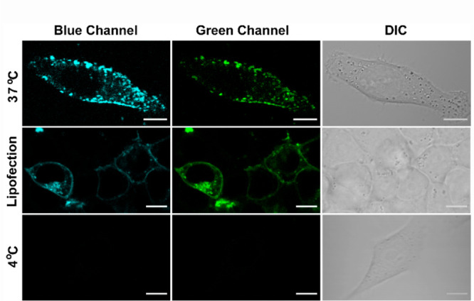
Figure 3.

Representative confocal single z-plane images of living HeLa cells. Cells were incubated with 7 μM DAN-APS for 15 min at either 37 °C (first row) or 4 °C (third row) in serum-free DMEM media, pH 7.4. For the lipofection experiment, DAN-APS (10 μg) was incorporated into live HeLa cells via lipofection (second row, 37 °C) and washed. DAN-APS was excited with a two-photon laser (λex: 780 nm). Fluorescence emission: blue channel (λem: 420–460 nm); top row green channel (λem: 560–590 nm); middle and bottom row green channel (λem: 510–540 nm). Differential interference contrast (DIC) images in right column. Scale bar, 10 μm.
