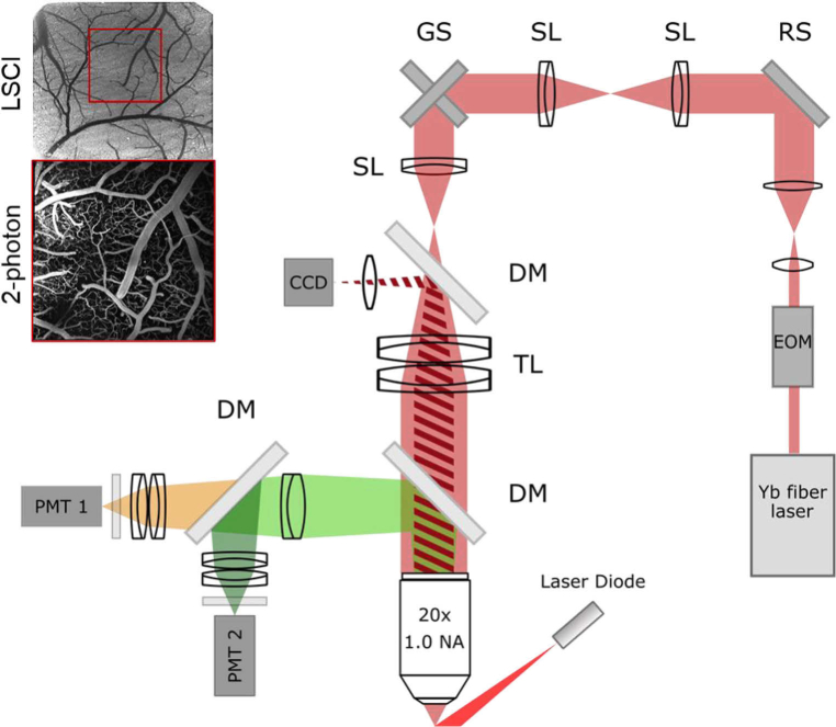
Fig. 1.
Resonant-galvo-galvo microscope schematic. EOM: electro-optic modulator. RS: resonant scanner (on a tip, tilt, and rotation stage). SL: scan lens (f = 50 mm). GS: galvanometer scanners. DM: dichroic mirror. TL: tube lens (f = 200 mm). PMT: photomultiplier tube. Excitation sources: Yb fiber laser (λ=1050 nm) for two-photon imaging, laser diode (λ=820 nm) for speckle imaging. Top left: sample LSCI and 2-photon images demonstrate use of LSCI image for informing 2-photon imaging location (red box).

