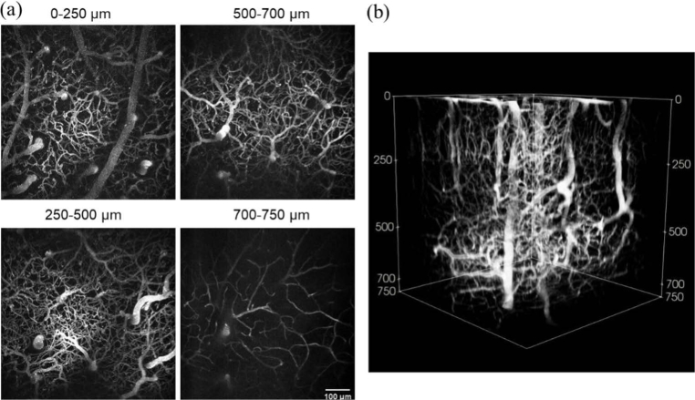
Fig. 3.
In vivo two-photon microscopy images of cortical vasculature taken with resonant-galvo scanning. (a) x-y maximum intensity projections showing frame averages of 15 frames from 0 to 250 µm, 40 frames from 250 to 500 µm, 100 frames from 500 to 700 µm, and 360 frames from 700 to 750 µm. (b) Three-dimensional reconstruction of the stack from which the projections in (a) were taken.

