Fig. 9.
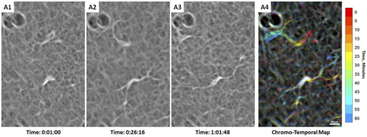
Two adjacent hyalocytes showing different movement imaged with AOSLO in a 31-year-old male ( Visualization 8 (15.2MB, mov) ). (A1-3). The lower cell appears more ramified with a spindle-like configuration whose cell body remains relatively stationary, as suggested by the white cell body color in the chromo-temporal map (A4), which is a composite of 13 images captured ∼5 minutes apart. A video of the ROI which includes these two cells is shown in Visualization 9 (19.3MB, mov) . In contrast, the upper cell has relatively shorter processes and appears more active with more substantial displacement and shape variation, as evidenced by the chromatic trail in the chromo-temporal map (A4). The dark circular features at the top left of each panel are likely microcysts, which are commonly found in sparse number throughout the retina. Time of acquisition of each image is displayed in hrs:mins:secs.

