Fig. 4.
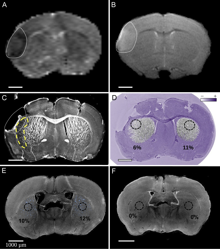
XPCT identifies subcortical white matter loss in mouse models of ischemic stroke and preterm birth. (A-D) MRI and XPCT data in a mouse model of ischemic stroke (mouse #1 of Table 2). (A) In vivo MRI data of mouse brain with pMCAO (single slice with thickness) imaged at 6 hours post-ischemia. The ischemic lesion (white dotted line) appears hypointense on apparent diffusion coefficient map (A) and hyperintense on T2-weighted image (B); (C) MIP over 107 slices ( ) of XPCT data obtained at the same slice level in the same mouse after sacrifice at day 9 post-ischemia. At this stage, the infarcted cortex tended to come off along the perfusion, extraction and dehydration steps of the brain. White matter loss is clearly seen in the subcortical peri-lesional area in this animal (yellow dashed line); (D) Segmentation of caudate putamen white-matter fibers and quantification of fiber density in symmetrical regions (percentage calculated in 3D ROIs over 100 slices). The background XPCT native image is represented in false colors to provide a cresyl violet-like contrast using a home-made colormap. (E-F) XPCT data in a mouse model of preterm birth: native (single axial slice) XPCT images in (E) normoxic and (F) hypoxic mice at post-natal day #11. White-matter fiber tracts were segmented and quantified in the striatum as shown in blue in normoxic animal (mouse #3 of Table 2) (E) but barely detectable in the animals that underwent neonatal chronic hypoxia (mouse #5 of Table 2) (F).

