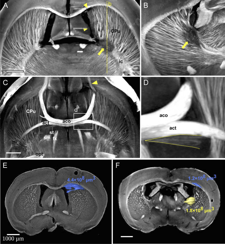
Fig. 5.
XPCT detects white-matter fiber tract damage in a mouse model of focal demyelination. (A) MIP of axial views over 100 slices ( ) at the level of the lateral ventricles. Focal demyelination is clearly seen in the internal capsule (arrow). Other focal lesions are shown in the ipsilateral (right) side with an arrow head such as lateral ventricle collapse; (B) MIP of sagittal views at the level of the lateral ventricle over 100 slices ( ); Again, focal demyelination is clearly seen in the internal capsule (arrow); (C) MIP of axial views over 100 slices ( ) at the level of the anterior commissure; degeneration of white-matter fiber tracts can be seen in the ipsilateral anterior part of the anterior commissure (arrowhead); (D) Disorganization of the ipsilateral stria terminalis is also seen compared to the contralateral stria, despite the small size of this fiber tract (dashed lines). (E-F) Segmentation and quantification of lesion volumes in two separate mice where demyelinated lesions were induced with the LPC model.

