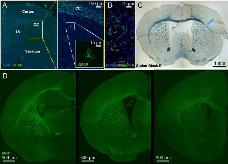
Fig. 7.
Brain preparation for XPCT does not prevent further brain evaluation with immunohistochemistry. (A) Constitutive expression of astrocytes GFAP in healthy mouse (CC: corpus callosum, LV: lateral ventricle); (B) Collagen IV overexpression in the ischemic lesion of a pMCAO mouse; (C) Sudan Black B staining (myelin marker) of a pMCAO mouse with demyelination of the corpus callosum (blue arrow) and (D) Myelin basic protein (MBP) staining in neonatal mice (P11); three slice levels are shown.

