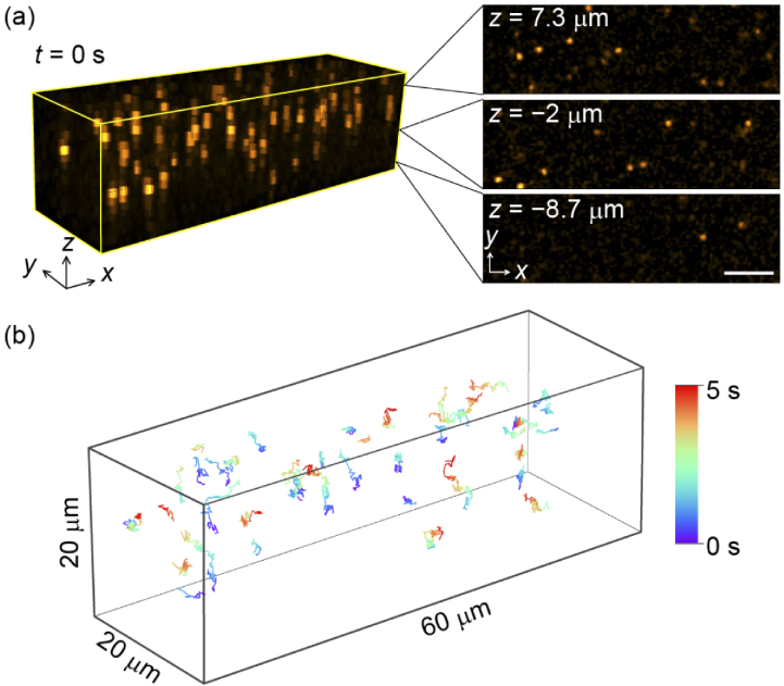
Fig. 4.
Video-rate imaging of 1-µm orange beads suspended in water with an acquisition rate of 31 vps. (a) 3D view of the image at t = 0. The rendered volume size is 60 × 17.8 × 20 µm3. The xy planes at z = 7.3, −2, and −8.7 µm are displayed on the right-hand side. The scale bar is 10 µm. (b) 3D trajectory of the Brownian motion of 44 beads recorded during 5 s. Only the beads wandering over 30 frames within the observing volume are depicted.

