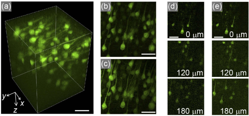
Fig. 6.
Acquisition of pyramidal neurons in a fixed H-line mouse brain with an acquisition volume of 200 × 200 × 260 µm3. (a) 3D view reconstructed from 13 volumes acquired by light needle scanning with a stack pitch of 20 µm (see Visualization 2 (7.5MB, mp4) ). (b) Maximum intensity projection image along the z axis of (a). (c) The corresponding image acquired using conventional Gaussian beam stacking with a z pitch of 1.33 µm, resulting in 196 images, for the same region. (d), (e) Representative xy planes at specific z positions, noted in each panel, for (b) and (c) are shown in (d) and (e), respectively. Each scale bar is 50 µm.

