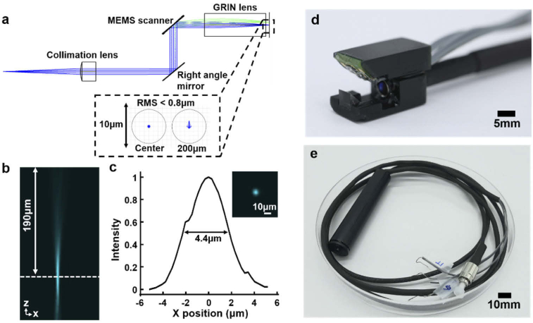
Fig. 4.
a) The calculated spot diagrams at the center and the scanning amplitude of 200 μm on the focal plane. The circles represent an airy disk. b) The measured beam focusing through the fully packaged confocal microscope catheter. c) The beam profile at the focal plane. The working distance is about 190 μm from the GRIN lens, and the beam spot is 4.4 μm at the focus. Optical images of d) the Lissajous scanning module and e) the fully packaged handheld confocal microscope catheter.

