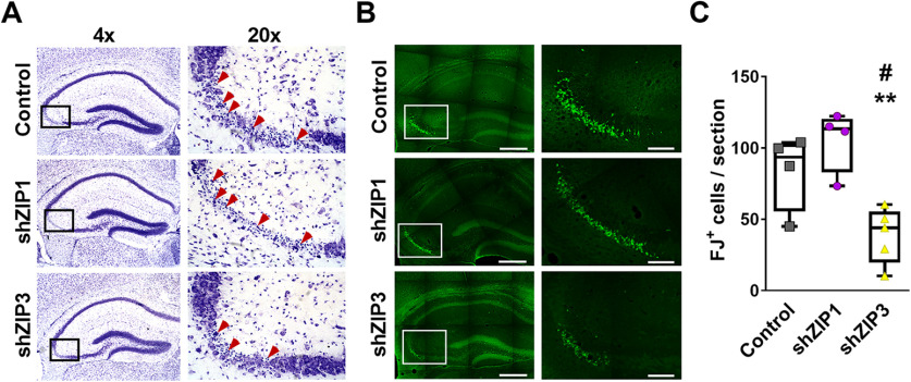
Figure 6.
Silencing of hippocampal ZIP3 expression prevents seizure-induced neurodegeneration of CA3 pyramidal neurons. A, Representative low (4×, left panel) and high (20×, right panel) magnification micrographs of Nissl staining showing neurodegeneration in the CA3 pyramidal layer, 24 h following kainic acid administration in vivo, leading to grade 5 seizures in mice transduced with shRNA-RFP-AAV control empty vectors (top panels) or vectors aimed to silence ZIP1 (middle panels) or ZIP3 (bottom panels). Arrows mark cells in which apoptotic bodies are recognized. B, Adjacent sections (to those presented in A) were stained with Fluoro-Jade B (green) marking degenerating neurons. Representative low (left, 4×) and high (right, 20×) magnification images are shown, as in A. Scale bar: 400 µm in 4× and 100 µm in 20×. C, Quantification of FJ+ CA3 pyramidal neurons (n = 4 mice for control and shZIP1, n = 5 mice for shZIP3; #p ≤ 0.05 compared with control; **p ≤ 0.01 compared with shZIP1, using ANOVA).

