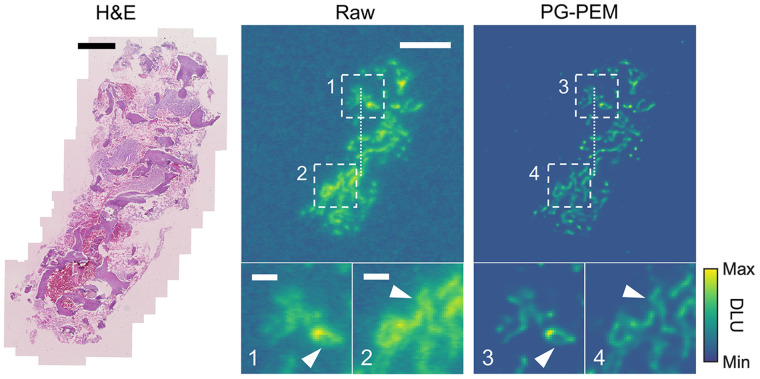
FIGURE 6.
PG-PEM restoration in α-particle radiotherapy specimens. From left to right: hematoxylin- and eosin-stained histologic image of bone biopsy sample from patient with 223RaCl2-treated metastatic castration-resistant prostate cancer, and corresponding raw and PG-PEM–restored DAR images. Scale bars: 1 mm (hematoxylin and eosin); 2.3 mm (raw); 0.5 mm (insets 1 and 2). DLU = digital light unit.

