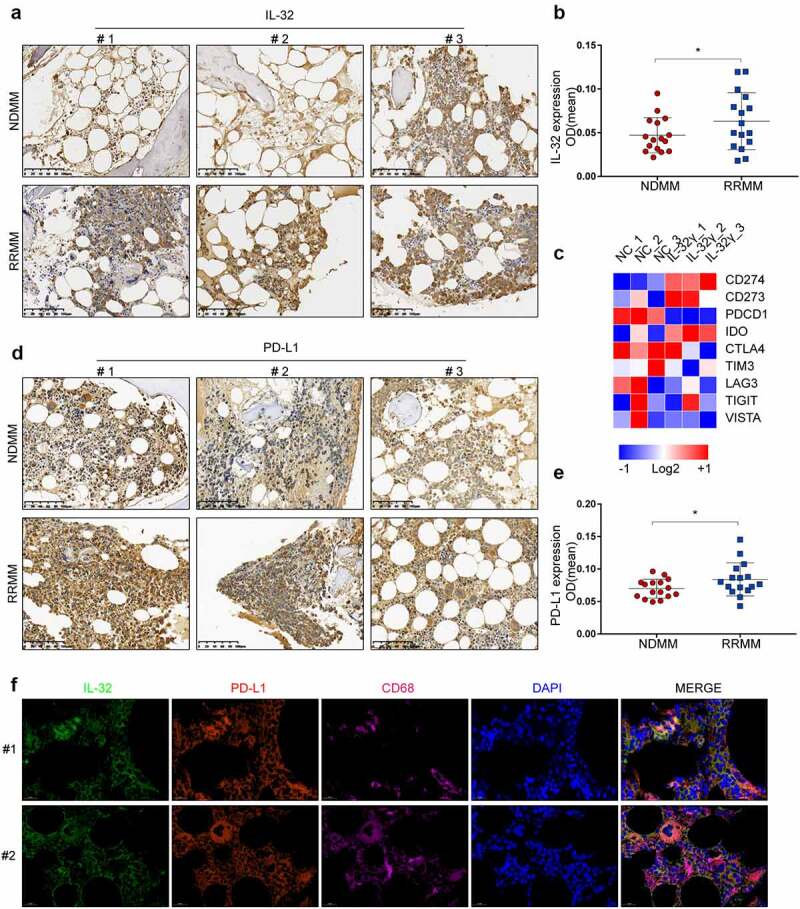
Figure 1.

IL-32 and PD-L1 are highly expressed in myeloma patients and correlated with disease stage. (a) Immunohistochemistry (IHC) analysis of BM biopsies from patients with NDMM and RRMM showing IL-32 expression. RRMM samples showed a higher IL-32 staining intensity compared to NDMM (scale bar, 100 μm). (b) The scores were determined for quantitative staining of IL-32 in patients with NDMM and RRMM. Results are expressed as mean ± SD (n = 16). (c) Macrophages generated from PBMCs of three independent, healthy donors were treated with IL-32γ (40 ng/ml) for 24 h. RNA was extracted for RNA sequencing. The heat map on immune checkpoint-related genes was shown. (d) Representative IHC images in BM biopsies from patients with NDMM and RRMM showing PD-L1 expression (Scale bar, 100 μm). (e) The scores were determined for quantitative staining of PD-L1 in patients with NDMM and RRMM. Results are expressed as mean ± SD (n = 16). (f) Immunofluorescence analysis of IL-32 (Green), PD-L1 (Red), and CD68 (Pink) expression in tumor tissues. Scale bar, 20 μm.
