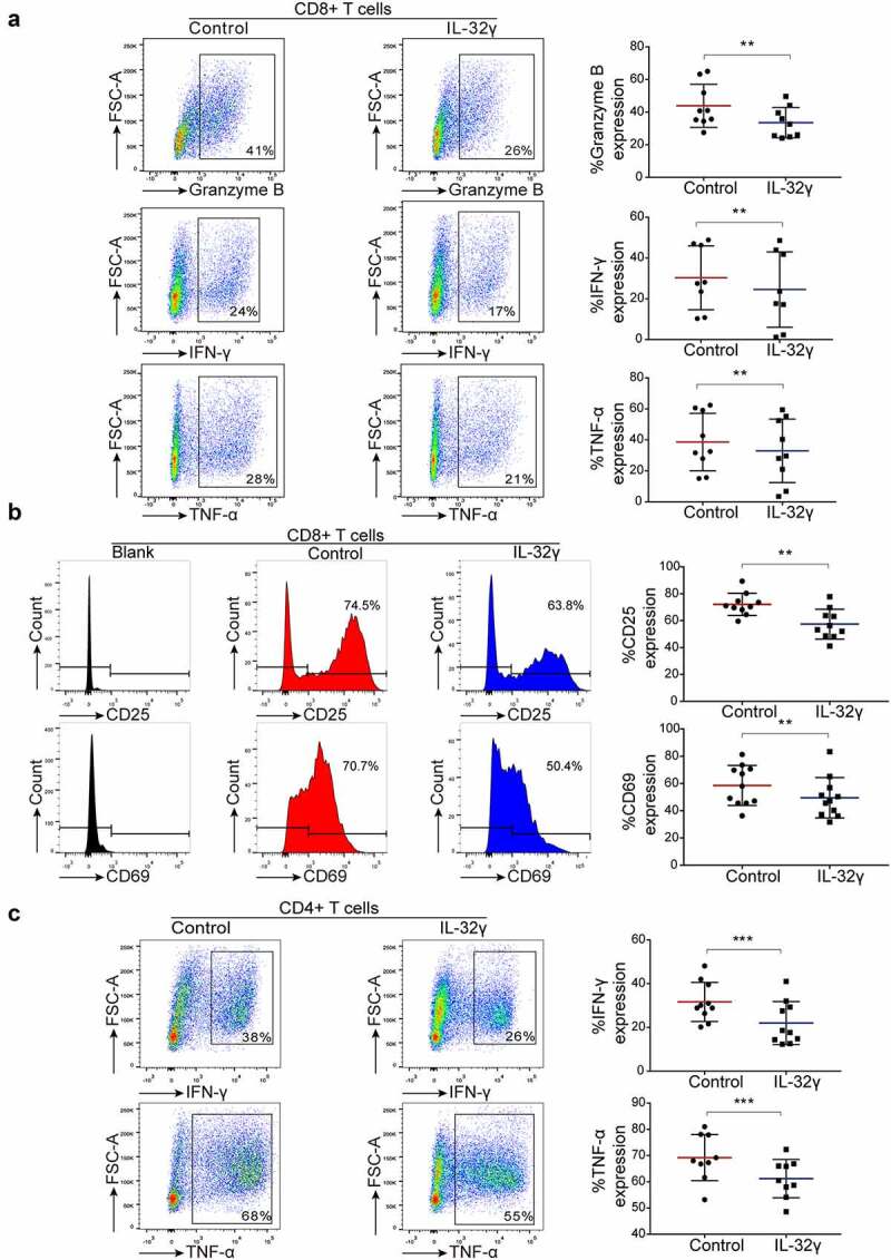
Figure 4.

IL-32γ treated macrophages inhibit T cell function and suppress T cell activation. (a) Macrophages were exposed to medium (Control) or IL-32γ (40 ng/ml) for 24 h and then co-cultured with healthy purified CD8 + T cells (macrophages: 35,000/well; T cells:35,0000/well). The levels of Granzyme B, IFN-γ, and TNF-α production were determined by flow cytometry after 48 h. (b) The expression of the activation markers CD25 and CD69 was measured by flow cytometry after 48 h. (c) Macrophages were exposed to medium (Control) or IL-32γ (40 ng/ml) for 24 h and then co-cultured with healthy purified CD4 + T cells (macrophages: 35,000/well; T cells: 35,0000/well). The levels of IFN-γ and TNF-α were determined by flow cytometry after 48 h. Data are presented as the mean ± SD of at least three independent experiments; *p < .05, **p < .01, ***p < .001.
