Figure 3.
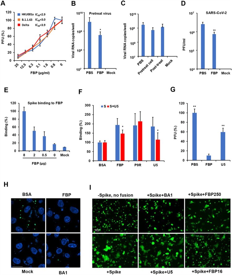
FBP inhibited SARS-CoV-2 infection in vitro. (A) Antiviral activity of FBP against SARS-CoV-2 in Vero-E6 cells (n = 3). SARS-CoV-2 variants were treated with FBP for cell infection. PFU (%) was normalized to the untreated viruses. (B) SARS-CoV-2 was treated with FBP (25 μg ml−1) and then infected cells for 1 h. Viral RNA copies in cell lysate were measured by RT-qPCR at 8 hpi (n = 5). (C) Cells were treated with FBP (25 μg ml−1) for 1 h before viral infection (Pretreat cell), and FBP was added to cells at 1 hpi (Post-treat). Viral RNA copies in cell lysate were measured by RT-qPCR at 8 hpi (n = 3). Mock, uninfected cells. (D) FBP inhibited SARS-CoV-2 by targeting the virus (n = 3). The virus (1 × 10⁶ PFU/ml) was treated by FBP (500 μg ml−1) and then was diluted to 1000 folds for the plaque assay. * indicates P < .05. P-values were calculated by the two-tailed Student’s t-test compared with PBS. (E) Dose-dependent binding of FBP to spike protein (n = 4). Spike binding to indicated FBP (8, 2 and 0.5 μg) and BSA (Mock) on the ELISA plate. Relative binding (%) was the OD values normalized to the OD value of FBP (8 μg). (F) FBP could bind to S protein, and U5 blocked the binding between FBP and S protein (n = 4). S protein of SARS-CoV-2 and S treated with U5 (S + U5) were added to the ELISA plate for binding to peptides coated on the ELISA plate. * indicates P < .05 compared with untreated S. (G) U5 (25 μg ml−1) showed weaker antiviral activity than that of FBP (25 μg ml−1) against SARS-CoV-2 (n = 4). The antiviral activity was measured by the plaque reduction assay. (H) FBP inhibited endosomal acidification. Vero-E6 cells were treated by BSA, FBP (25 μg/ml) or bafilomycin A1 (BA1, 25nM). The low pH indicator (green) showed the low pH in endosomes. Nuclei were stained by DAPI (blue). Untreated cells (Mock) were the negative control. Scale bar = 20 μm. Experiments were repeated twice. (I) FBP inhibited spike-ACE2-mediated cell–cell fusion. Co-cultured cells (293T/spike and 293T/ACE2 cells) were treated with FBP (250 and 16 μg ml−1), U5 (250 μg ml−1) and bafilomycin A1(BA1, 50 nM). Cells without spike (-spike) were served as the no-fusion control. Scale bar = 100 μm. Experiments were repeated twice.

