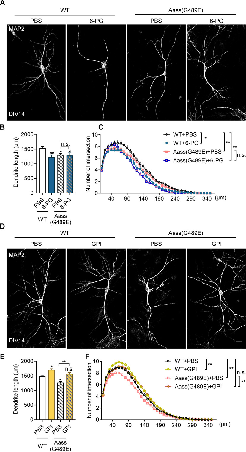
Figure 11.
Supplementation of GPI rescues defective dendritic development caused by SDH mutation. A, Representative images of primary neurons dissected from WT or Aass (G489E) brains. The neurons treated with or without 6PG (50 μm) from DIV7 to DIV14, and stained with MAP2 antibodies at DIV14. Scale bars: 20 μm. B, C, Quantitative analysis of the total basal dendritic length (B) and the basal dendritic complexity (C) of cultured neurons in A. B, One-way ANVOVA, F(3,11) = 0.9085. WT + PBS versus WT + 6PG, **p = 0.01; WT + PBS versus Aass (G489E) + PBS, *p = 0.0355; WT + PBS versus Aass (G489E) + 6 PG, *p = 0.0115; Aass (G489E) + PBS versus Aass (G489E) + 6 PG, p = 0.9041. C, Univariate ANOVA, WT + PBS versus WT + 6PG, F(1,4) = 19.287, *p = 0.012; WT + PBS versus Aass (G489E) + PBS, F(1,5) = 21.306,**p = 0.006; WT + PBS versus Aass (G489E) + 6 PG, F(1,5) = 16.737, **p = 0.01; Aass (G489E) + PBS versus Aass (G489E) + 6 PG, F(1,6) = 5.647, p = 0.055. WT + PBS, 104 neurons, n = 3 brains; WT + 6PG, 58 neurons, n = 3 brains; Aass (G489E) + PBS, 94 neurons n = 4 brains; Aass (G489E) + 6PG, 40 neurons, n = 4 brains. D, Representative images of primary neurons dissected from WT or Aass (G489E) brains. The neurons treated with or without GPI (100 ng) from DIV7 to DIV14, and stained with MAP2 antibodies at DIV14. Scale bars: 20 μm. E, F, Quantitative analysis of the total basal dendritic length (E) and the basal dendritic complexity (F) of cultured neurons in D. E, One-way ANVOVA, F(3,12) = 1.189. WT + PBS versus WT + GPI, *p = 0.038; WT + PBS versus Aass (G489E) + PBS, *p = 0.0224; WT + PBS versus Aass (G489E) + GPI, p = 0.9360; Aass (G489E) + PBS versus Aass (G489E) + GPI, **p = 0.0081. F, Univariate ANOVA, WT + PBS versus WT + GPI, F(1,6) = 17.738, **p = 0.006; WT + PBS versus Aass (G489E) + PBS, F(1,7) = 19.996,**p = 0.003; WT + PBS versus Aass (G489E) + GPI, F(1,7) = 0.740, p = 0.418; Aass (G489E) + PBS versus Aass (G489E) + GPI, F(1,8) = 11.224, **p = 0.01. WT + PBS, 168 neurons, n = 4 brains; WT + GPI, 232 neurons, n = 4 brains; Aass (G489E) + PBS, 207 neurons, n = 5 brains; Aass (G489E) + GPI, 177 neurons, n = 5 brains. Data are presented as mean ± SEM; n.s., no significance, p > 0.05, *p < 0.05, **p < 0.01.

