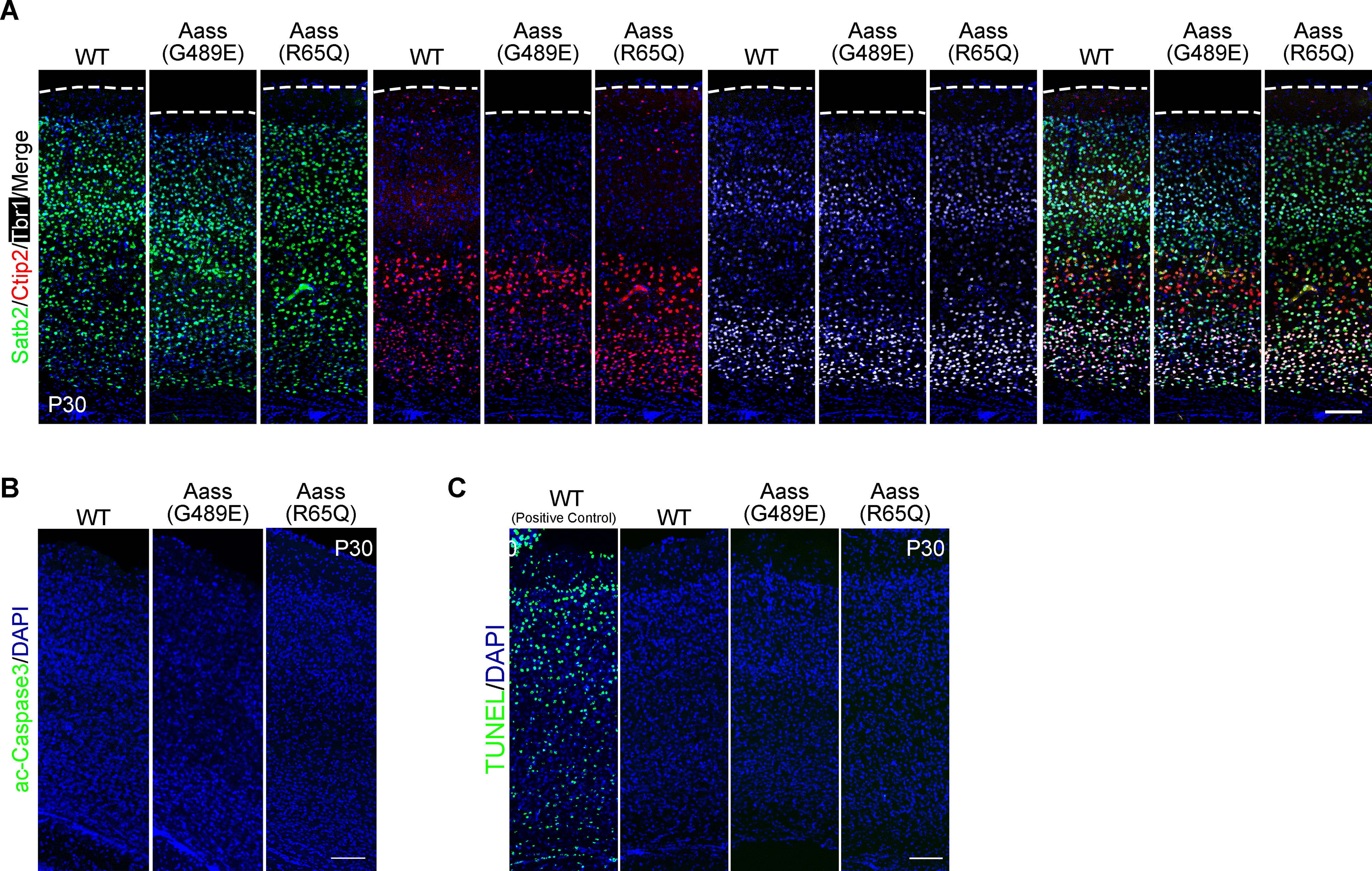
Figure 2.

Mutations in LKR or SDH do not affect brain lamination and cell survival. A, Immunostaining images of coronal sections of WT, Aass (G489E), and Aass (R65Q) brains at P30. Different markers were used to label the cortex layers. Satb2, projection neuron marker (green); Ctip2, Layer V (red); Tbr1, Layer VI (white). Nuclei were stained with DAPI (blue). Scale bars: 100 μm. B, Coronal sections of WT, Aass (G489E), and Aass (R65Q) brains stained with active-Caspase 3 (green) at P30. Nuclei were stained with DAPI (blue). Scale bars: 100 μm. C, Cell death detection using TUNEL staining (green) in the coronal sections of WT, Aass (G489E), and Aass (R65Q) brains at P30. Nuclei were stained with DAPI (blue). Scale bars: 100 μm.
