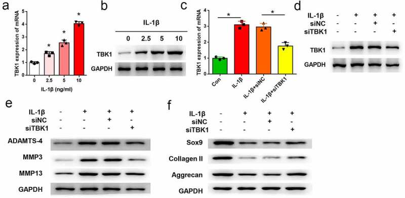
Figure 3.

Silencing of TBK1 inhibited ECM degradation in OA cell model. ATDC5 cells were treated with various doses of IL-1β for 12 h, after which (a) mRNA and (b) protein levels of TBK1 were analyzed by qRT-PCR and Western blotting, respectively. ATDC5 cells were transfected with siTBK1 or siNC and then stimulated with 10 ng/ml IL-1β for 12 h. (c) mRNA and (d) protein levels of TBK1 were detected by qRT-PCR and Western blotting, respectively.(e) matrix-degrading enzymes (ADAMTS-4, MMP3, MMP13), and (f) ECM-related molecules (SOX9, collagen II, aggrecan) were analyzed by Western blotting. *p < 0.05.
