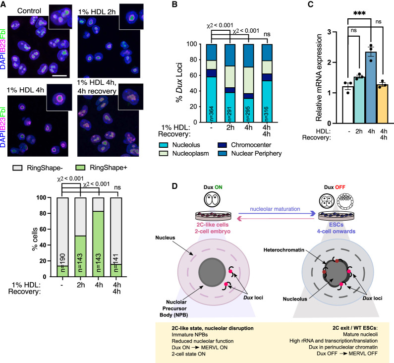
Figure 6.
Disruption of LLPS induces Dux movement and activation. (A) Immunofluorescence for nucleolar markers B23 and Fbl after the indicated times of incubation with 1% 1,6-hexanediol (HDL) with or without washout and recovery in normal media, and quantification of the percentage of cells with RingShape+ nucleoli (below). Scale bar, 20 µm. (n) Number of cells. P-values, χ2 test with Bonferroni adjustment for multiple comparisons. (B) Scoring of Dux locus nuclear positioning following HDL treatments from Dux immuno-FISH experiments. (n) Number of nuclei scored from two FISH experiments. P-values, χ2 test, with Bonferroni adjustment for multiple comparisons. (C) Expression of Dux by qRT-PCR following HDL treatment. Data are mean ± SEM for n = 3 biological replicates, representative of two independent experiments. P-values, one-way ANOVA with Dunnett correction for multiple comparisons. (D) Model: Nucleolar maturation allows for Dux repression and two-cell exit. In early embryos and 2C-like cells, NPBs have altered morphology, reduced function, and reduced chromatin association. We propose that this provides a permissive environment for Dux and subsequent 2C/MERVL expression. In mature nucleoli with high rRNA output, Dux is recruited to perinucleolar chromatin and is repressed. Disruption of nucleolar integrity via iPol I or inhibition of nucleolar phase separation releases Dux and leads to its derepression.

