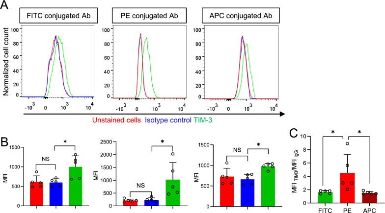
Fig. 3.
PE-conjugated antibody outperforms FITC and APC conjugated antibodies in detecting microglia TIM-3. A Distribution of fluorescence intensities from nontreated microglia, FITC, PE, and APC conjugated TIM-3 antibodies treated, and their respective isotype control antibody-treated microglia. B The MFI of microglia treated as in (A). C Comparison of MFI TIM-3/MFI IgG of FITC, PE, and APC conjugated TIM-3 antibodies (n = 5 mice). Bars represent means ± SD. Comparisons were made by one-way ANOVA test with Tukey’s post hoc multiple comparisons test. *P < 0.05; **P < 0.01; NS, not significantly different

