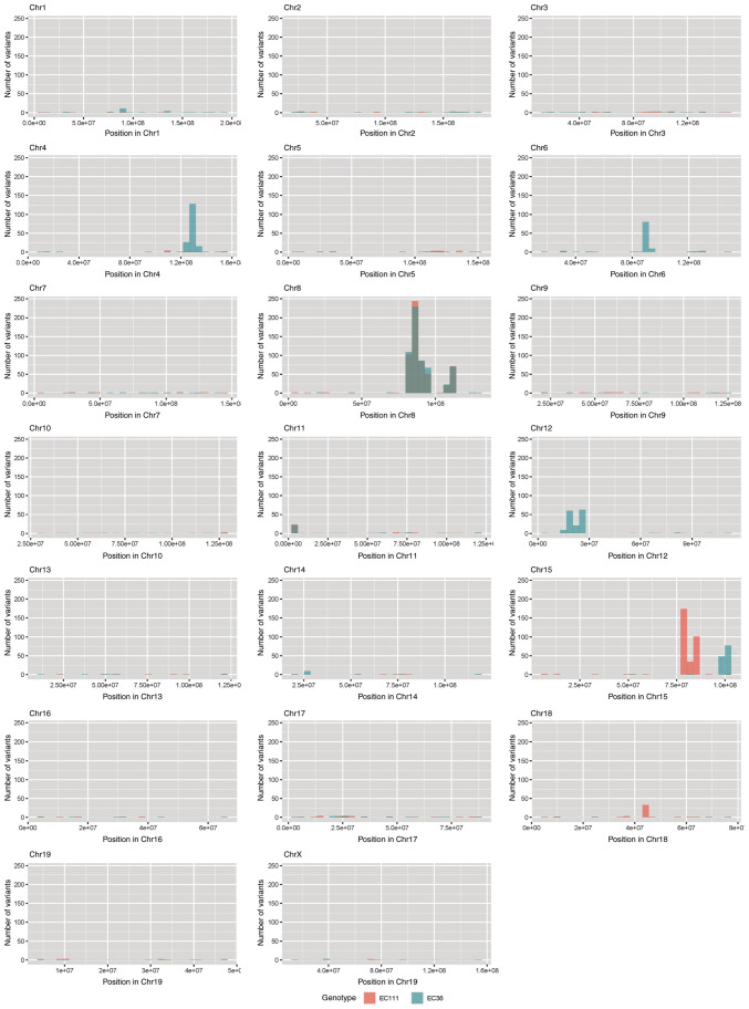
Figure 2.
Somatic mutation analysis. Bar plots showing the number of nucleotide variants found in both mCyld−/− cell lines but not in mCyld+/+ cell lines. Each bar represents the number of variants per 10 million base pairs along the chromosomal location in mCyld−/− cell line #1 (red) and #2 (blue). Common variants found in both mCyld−/− cell lines are only present on Chr 8 (gray) due to knockout of Cyld. Chr, chromosome; CYLD, CYLD lysine 63 deubiquitinase.

