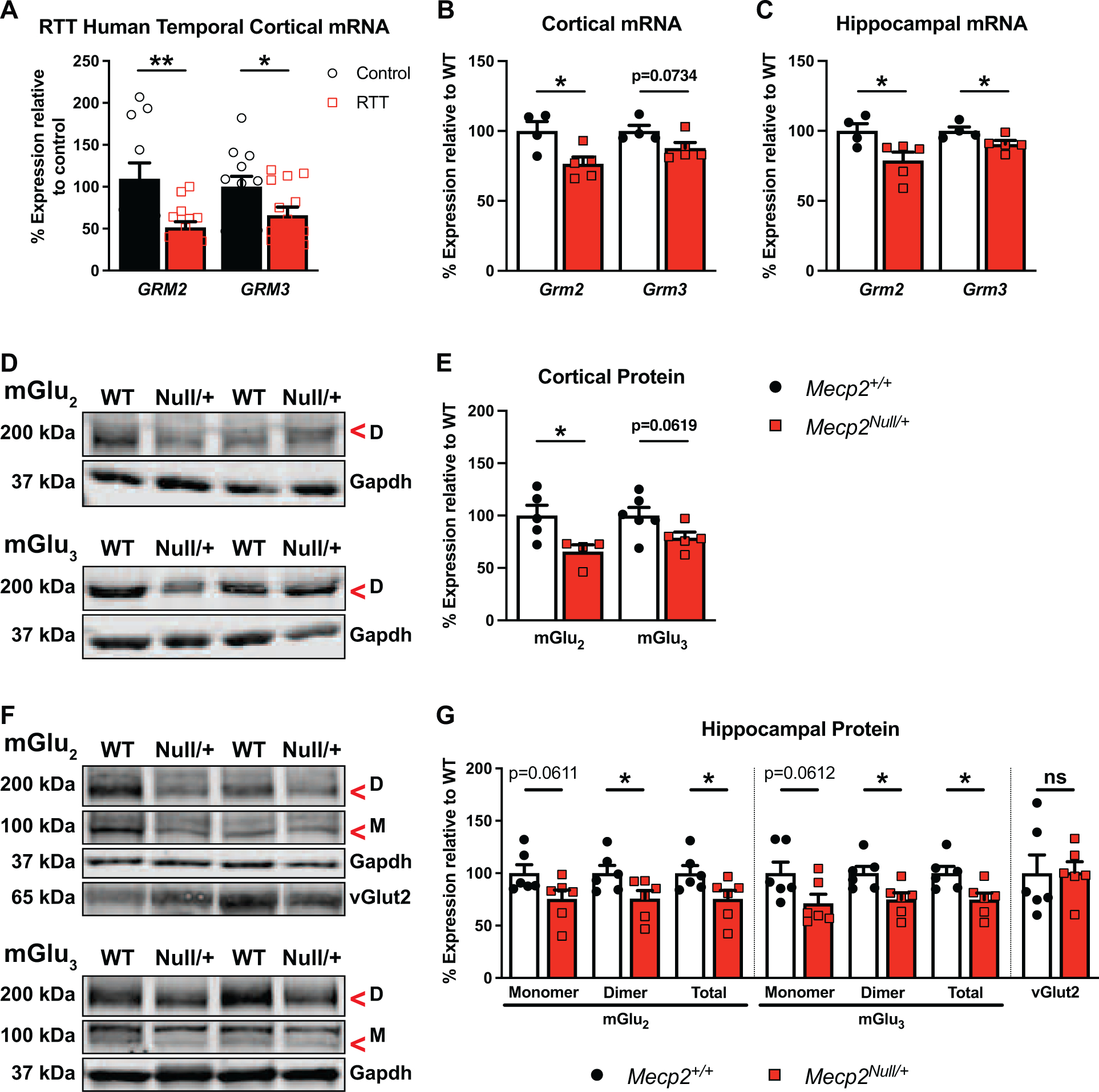
Figure 1: mGlu2 and mGlu3 expression is decreased in RTT patients and mice.

(A) Compared to controls (black bars / white circles, n=11–12), GRM2 and GRM3 mRNA are both decreased in RTT patients in temporal cortex autopsy samples (red bars / white squares, n=14) (GRM2: t(23)=3.144, p=0.0045; GRM3: t(24)=2.196, p=0.0380). RTT patient samples used in this study have truncated MeCP2 mutations, specifically R168X, R255X and R270X (see Supplementary Table 1). (B-C) Compared to littermate controls, Mecp2+/+ (white bars / black circles, n=4–5), Grm2 and/or Grm3 mRNA levels are decreased in 20–25-week-old Mecp2Null/+ mice (red bars / squares, n=4–5) (cortex: Grm2: t(7)=2.897, p=0.0231; Grm3: t(7)=2.104, p=0.0734; hippocampus: Grm2: t(7)=2.599, p=0.0355; Grm3: t(7)=2.460, p=0.0435). (D) Representative immunoblots illustrating cortical dimeric (“D”, 200 kDa) form of mGlu2 or mGlu3 and Gapdh (37 kDa) loading control in Mecp2+/+ (“WT”) and Mecp2Null/+ (“Null/+”) animals. (E) Cortical mGlu2 protein expression is significantly decreased in Mecp2Null/+ mice (n=4–5) relative to Mecp2+/+ animals (n=5–6) (mGlu2: t(7)=2.715, p=0.0300; mGlu3: t(9)=2.131, p=0.0619). (F) Representative immunoblots for hippocampal proteins as in (D) with the addition of the mGlu2 or mGlu3 monomeric protein (“M”, 100 kDa) and vGlut2 (65 kDa). (G) Compared to Mecp2+/+ animals (n=5–6), mGlu2 and mGlu3 proteins (total = monomer + dimer) are reduced in the hippocampus of Mecp2Null/+ mice (n=5–6) (mGlu2: Monomer: t(10)=2.109, p=0.0611; Dimer: t(10)=2.285, p=0.0454; Total: t(10)=2.250, p=0.0482; mGlu3: Monomer: t(10)=2.108, p=0.0612; Dimer: t(10)=2.798, p=0.0188; Total: t(10)=2.811, p=0.0185). vGlut2 is unchanged between genotypes (t(10)=0.04986, p=0.9612). Student’s t-test. ns (not significant), *p<0.05, **p<0.01.
