Figure 4.

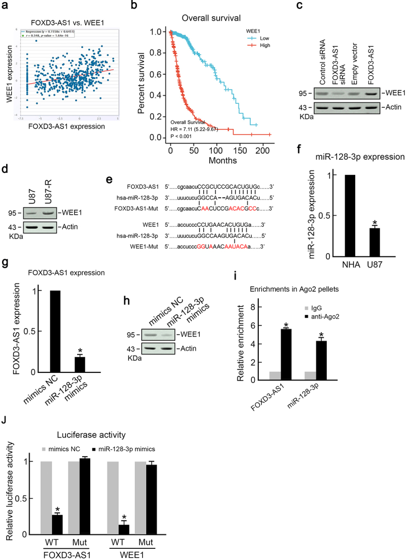
FOXD3-AS1 promoted WEE1 expression by sponging miR-128-3p.
(A) Positive correlation of FOXD3-AS1 and WEE1 in GBM patients. Data from TCGA database. (B) Kaplan–Meier analysis results showed that higher levels of WEE1 were associated with poorer prognosis of GBM patients. (C) After transfection using specific reagents, immunoblotting was conducted to determine WEE1 expression in U87 cells, with actin as a loading reference. (D) The WEE1 level was elevated in U87-R cells compared to the parental U87 cells, with actin as a loading reference. (E) Sketch map showing possible binding sites of miR-128-3p in WEE1 and FOXD3-AS1.(F) MiR-128-3p level was elevated in U87 cells compared to healthy NHA cells (N = 3, *p < 0.05). (G) Overexpression of miR-128-3p reduced FOXD3-AS1 level in U87 cells (N = 3, *p < 0.05). (H) Overexpression of miR-128-3p suppressed WEE1 expression in U87 cells. Notably, actin served as the loading control. (I) Relative abundances of miR-128-3p and FOXD3-AS1 in the RISC complex detected using the RIP assay with the use of anti-Ago2 antibody (N = 3, *p < 0.05). (J) Dual-luciferase reporter assay. Specific reagents were used to transfect U87 cells for 48 h, and then the relative luciferase activities were measured using the Dual-Luciferase Reporter assay system (Promega) (N = 3, *p < 0.05).
