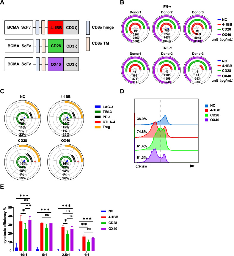
Fig. 1.
Characterizations of antitumour efficacy among CD28, OX40 and 41BB based CAR-T cells. A The structure of the BCMA-CAR consists of the same variable region of the BCMA single-chain antibody, the CD8 hinge and transmembrane regions, different costimulatory molecules (from 41BB, CD28, or OX40) and CD3ζ. B Coincubation of effector cells with target 8226 cells for 24 h at a ratio of 5:1 between D10 and D15 (different days were used in different donors) and the supernatant was collected. Cytokines were detected by a human Th1/Th2/Th17 kit using flow cytometry. The qualitative analysis of the expression of IFN-γ, TNF-α was performed with Phyton 3.7 using the Matplotlib package (https://matplotlib.org/) (n = 3 donors). C The expression of the exhaustion-related markers LAG-3, TIM-3, PD-1, and CTLA-4 on T cells expressing BCMA-CAR were measured on day 7 (D7) (n = 3 donors). Data were analyzed with FlowJo software, and graphs were plotted with Phyton 3.7 using the Matplotlib package (https://matplotlib.org/). D The Cell Trace TM CFSE Cell Proliferation Kit was used to detect cell proliferation. On D13, effector T cells were stained with CFSE (CFDA-SE) dye and incubated with target K562 (negative control) and 8226 cells at a ratio of 5:1. After 5 days of incubation, CFSE fluorescence intensity was detected by flow cytometry (K562 data not shown). E Effector T cells were incubated for 24 h with target K562 (negative control) and 8226 cells at E:T ratios of 10:1, 5:1, 2.5:1, and 1:1 on D13. Cytotoxicity was determined from the amount of released LDH in the culture supernatants using an LDH kit at a wavelength of 490 nm. The figure shows the result of effector T cells incubated with the 8226 target cells (n = 3, P < 0.001 and P < 0.01, error bars denote standard deviation)

