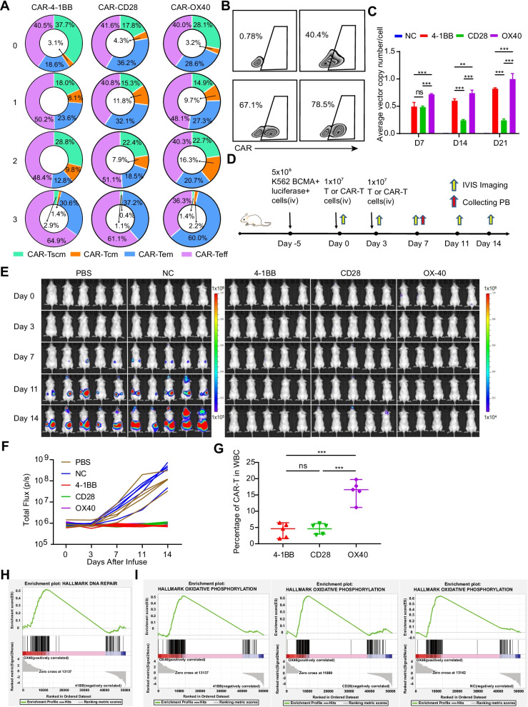
Fig. 2.
OX40-CAR-T cells are the least exhausted and have superior persistence. A Effector cells were subjected to three consecutive repeated stimulations with 8226 target cells (CAR+ cells and 8226 target cells at a ratio of 1:1; 3-day interval was used for each stimulation), changes in CAR+ cell subtypes were determined with flow cytometry (n = 3). B CAR+ cells from different experimental groups after repeated stimulation. As described in A, FlowJo was used for data analysis and presentation (n = 3). C The average copy number of the CAR gene in single cells was detected by qPCR technology on day 7 (D7), D14, and D21 after the cells were activated with anti-CD3/CD28 antibodies (n = 3, P < 0.001 and P < 0.01, respectively. error bars denote standard deviation). D Schematic outline of the mouse model experiment (n = 5). All the Methods and Materials were described in the Additional file 4. E Tumour progression was monitored by IVIS imaging. In order to scientifically show the small gap between different CAR-T, the scales are normalized for PBS and NC group with 1 × 10⁵ ~ 1 × 10⁶, and 4-1BB, CD28 and OX40 CAR-T group with 1 × 10⁴ ~ 1 × 10⁵. F Tumour progression was monitored by total flux of each mice. G The proportion of CAR-T in WBC (white blood cell) were detected from mice PB by flow cytometry on day 7. ***P ≤ 0.001, NS no significant. H Representative GSEA of DNA repair pathways and MSigDB hallmark gene set for two different BCMA-targeted CAR-T cells (OX40-CAR-T cells and 41BB-CAR-T cells). I Representative GSEA of the oxidative phosphorylation pathway and MSigDB hallmark gene sets for different BCMA-targeted CAR-T cells

