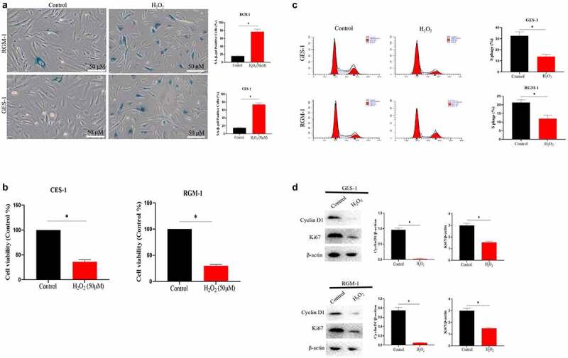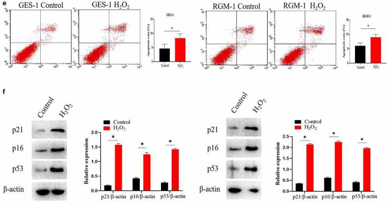
Figure 1.

(a). Sa-β-gal positive cells significantly increased by H2O2 stimulation. The experiments were performed by β-Galactosidase Staining Kit (Solarbio, G8860) according to the manufacturer’s instructions. (b). Cell proliferation were reduced after H2O2 treatment by MTT assay. GES-1 and RGM-1 cells were exposed to H2O2 for 2 h. The cells were then distributed into a 96-well cell culture plate (2 × 10⁴/well), and incubated for 24 h. After washing with PBS, the MTT (50 μL/well) reagent was added into each well and incubated for 120 min. The optical density (OD) value in the each well was detected with a microplate reader (Bio-rad, imark). n = 3 biological replicates. (c). The ratio of S phase cells were significantly decreased by H2O2 treatment. n = 3 biological replicates. (d). The expressions of cyclin D1 and Ki67 were also significantly down-regulated by Western-blot analysis. E. Analysis of cell apoptosis by flow cytometry. F. P15 and P16 were significantly up-regulated by H2O2 treatment. The data are shown as means ± SEM. Asterisks indicate significant differences (P < 0.05).
