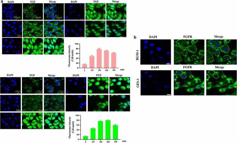
Figure 2.

(a). The GES-1 and RGM-1 cells were seeded on 6-well cell culture plates. 40% confluent cells were starved for 10 h. The cells were then stimulated with FITC-FGF for the indicated time points. After the cells were fixed, the cells were observed using CLSM. (b). FGF could not transport into the nucleus in the senescent cells. The average fluorescence intensity of 50 cells was measured and analyzed. Asterisks indicate significant differences (P < 0.05).
