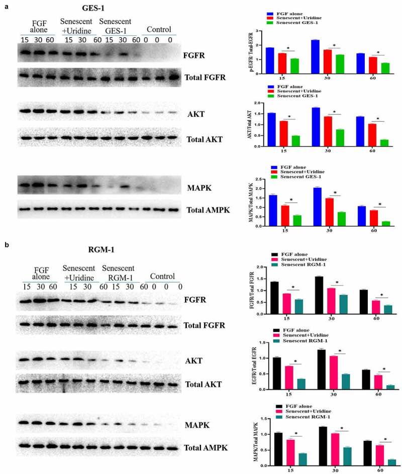
Figure 5.

Uridine treatment partially restored the signaling ability of FGF. GES-1 and RGM-1 cell were stimulated with FGF (30 ng/ml) for the indicated time point. Then, the cell lysate was used to lyse the cells, and then the cell lysate was centrifuged at 10,000 RPM/min for 10 min. the precipitate was discarded and the supernatant was collected. The cell concentration was measured by BCA method. The samples were separated by SDS-PAGE and transferred to PVDF membrane. After washing twice, the membrane was sealed with 5% BSA at 37°C for 2 h. After washing, the primary antibody was added and incubated for 12 h at 4°C. After washing, the secondary antibody was added and incubated for 2 h. After washing, the immunoprotein bands were detected by ECL kit.
