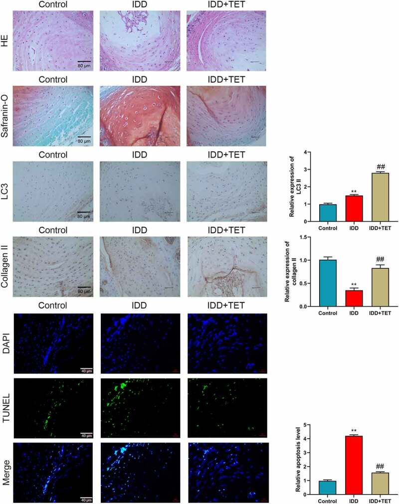
Figure 6.

TET delayed rat intervertebral disc degeneration induced by puncture in annulus fibrosus. (a-b) The pathological changes of intervertebral disc were detected by HE and Safranin-O staining. (c-f) LC3 and Collagen II expression in intervertebral disc were measured by immunohistochemistry, and the relative expression were showed in histograms. (g) The apoptosis was assessed by TUNEL assay. (h) Relative apoptosis level was showed in a histogram. **p < 0.01 vs. control group, ##p < 0.01 vs. TBHP group.
