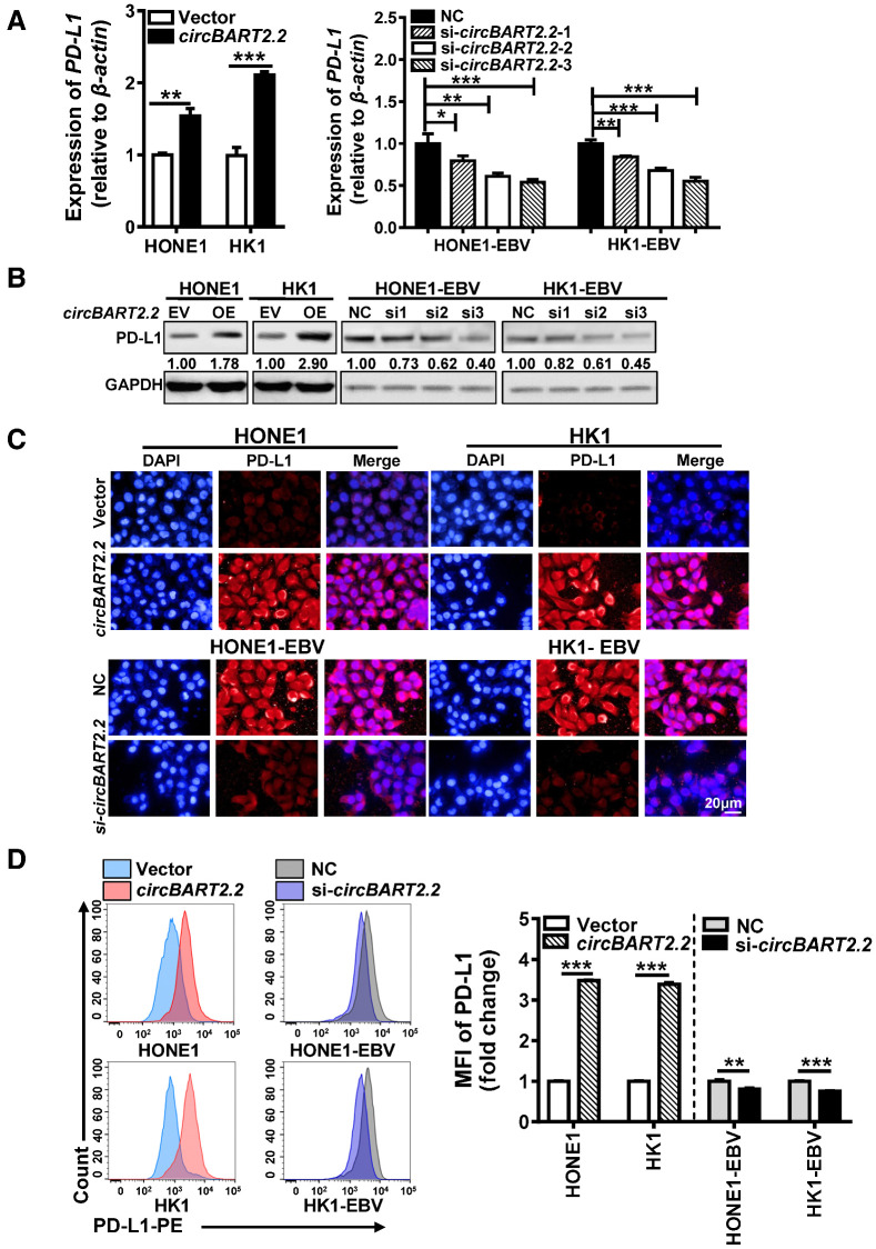
Figure 2.
circBART2.2 significantly upregulated PD-L1 expression. A, The mRNA expression of PD-L1 was examined in HONE1 and HK1 cells after circBART2.2 overexpression or in HONE1-EBV and HK1-EBV cells transfected with three circBART2.2 siRNAs, respectively, as demonstrated by RT-PCR. *, P < 0.05; **, P < 0.01; ***, P < 0.001. B, The expression of PD-L1 protein was examined in HONE1 and HK1 cells after circBART2.2 overexpression or in HONE1-EBV and HK1-EBV cells transfected with three circBART2.2 siRNAs, respectively, as demonstrated by Western blotting. C, PD-L1 expression (red) was measured in EBV-negative or -positive NPC cells after circBART2.2 overexpression or knockdown by immunofluorescence using anti–PD-L1 antibody. Nuclei were stained with DAPI (blue). Magnification, ×400. Scale bar, 20 μm. D, PD-L1 expression was measured in EBV-negative or -positive NPC cells after circBART2.2 overexpression or knockdown using flow-cytometric analysis using APC-stained anti–PD-L1 antibody. Left, original flow cytometry results; right, statistical results. Three independent replicates were evaluated for each group. MFI: mean fluorescence intensity. **, P < 0.01; ***, P < 0.001.

