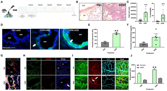
Figure 1.
Periostin expression is increased following chronic aeroallergen exposure in mice. (A) Schematic diagram of HDM-induced allergic airway inflammation in mice. (B) Female C57/Bl6 mice (6–8 weeks old) were exposed to either sterile PBS (10 μl intranasally) or house dust mite extract (HDM; 25 μg in 10 μL) 5 days a week for five consecutive weeks. Hematoxylin and eosin stained lung sections from PBS and HDM-exposed mice. (C) Bronchoalveolar lavage fluid total cell counts and percentage of eosinophils. (D) At the end of the allergen exposure protocol, lung sections obtained from PBS control and HDM-exposed mice were stained with an anti-periostin antibody (green) and DAPI to stain nuclei (blue). Images were taken at 400x and 630x magnification as stated. Arrows indicate periostin positive cells. (E) Expression of periostin was calculated in the area of interest around each airway using ImageJ. **p < 0.01, n = 3 representative of two independent experiments. (F) Bronchoalveolar lavage samples were collected and the periostin content was assessed by ELISA. n = 4 representative of two independent experiments. (G,H) Lung sections were stained for periostin (green), the pericyte marker PDGFRβ (red), and cell nuclei (DAPI, blue) to demonstrate the presence of periostin-expressing pericytes around the airways and blood vessels of HDM-exposed mice. (I) Tracheobronchial whole mounts were stained for the mesenchymal cell marker α-smooth muscle actin (α-SMA; red), the endothelial cell marker CD31 (cyan), and periostin (green) and imaged at 400x magnification; arrow indicate periostin-positive pericytes. (J) The expression of periostin and α-SMA in tracheobronchial whole mounts was calculated using ImageJ. AW, airway; BV, blood vessel. **p < 0.01, n = 3–5 representative of two independent experiments.

