Figure 2.
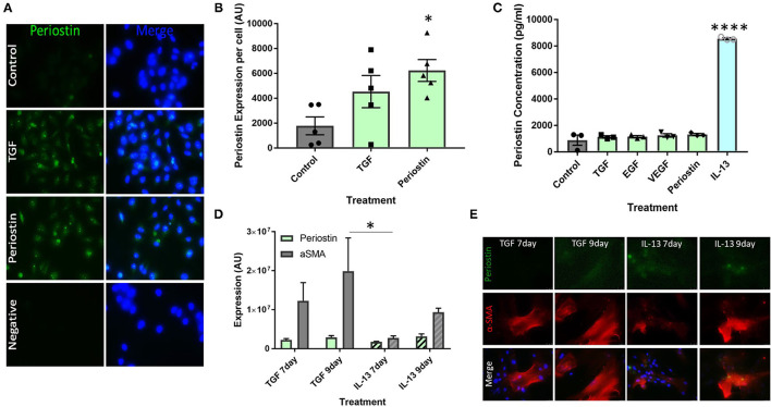
Type 2 inflammatory mediators stimulate periostin expression by pericytes. (A,B) Immunostaining performed on pericytes grown in pericyte medium and treated with 10 ng/ml of TGF-β or 100 ng/ml of periostin for 24 h. Cells were stained with an anti-periostin antibody (green) and the nuclear stain DAPI (blue). Images were taken at 400x magnification and intensity of periostin stain was calculated with ImageJ. Intensity of stain per field of view was divided by the number of cells in order to determine periostin expression per cell. *p < 0.05, n = 5 representative of two independent experiments. (C) Cultured pericytes were treated with 10 ng/ml TGF-β, 10 ng/ml EGF, 10 ng/ml VEGF, 100 ng/ml IL-13 or 100 ng/ml Periostin in pericyte medium for 7 days before the supernatant was harvested. The periostin content was assessed using an anti-periostin ELISA kit. ****p < 0.0001, n = 3 representative of two independent experiments. (D,E) Cultured pericytes were treated with 10 ng/ml TGF-β or 100 ng/ml IL-13 for either 7 or 9 days. Cells were stained with an anti-periostin antibody and an anti-αSMA antibody. The images were taken at 400x magnification and quantifications were made using ImageJ. *p < 0.05, n = 3–4 representative of two independent experiments.

