Figure 4.
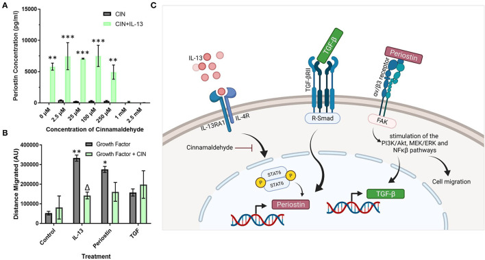
Cinnamaldehyde treatment suppresses periostin expression and pericyte migration. (A) Cultured pericytes were treated with 100 ng/ml IL-13 in pericyte media for 7 days with the addition of cinnamaldehyde for the last 3 days at the concentration stated in (A) or only cinnamaldehyde at the stated concentration for 3 days. The supernatants were harvested and the periostin content was assessed by ELISA. **p < 0.01, ***p < 0.001 vs. the same cinnamaldehyde concentration without IL-13, n = 2 representative of two independent experiments. (B) Scratch assay performed on pericytes that were grown in pericyte medium and treated with 10 ng/ml of TGF-β, 100 ng/ml of IL-13 or 100 ng/ml of periostin for 7 days with 1 mM cinnamaldehyde added for the last 3 days. Cells were transferred into media lacking serum and a scratch was made in each monolayer using a p200 pipette tip. Images were taken at 100x magnification immediately after scratching and 24 h later. The scratch width was determined using ImageJ and the average distance of cell migration was calculated. n = 3 representative of two independent experiments, Δ = p < 0.05 vs. the same growth factor treatment without cinnamaldehyde, *p < 0.05, **p < 0.01 vs. control treatment without cinnamaldehyde. (C) Schematic diagram of the putative signaling pathways regulating periostin expression in pericytes (created using Biorender).

