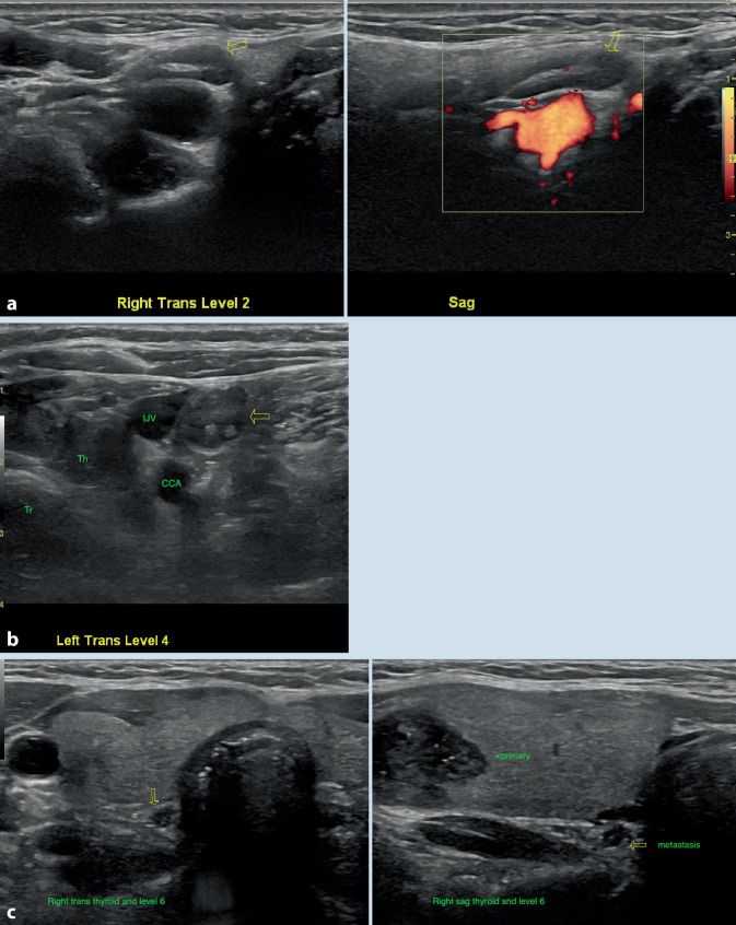
Fig. 6.
a Benign ovoid level 2 lymph node with central hyperechoic linear hilum (left panel, transverse view) and minimal superimposed hilar flow on power Doppler (right panel, sagittal view). b By contrast, rounded lymph node with metastatic papillary thyroid carcinoma, lacking visible hilum, with irregular margins and internal echogenic material (arrow). Tr trachea, Th thyroid with primary tumor, CCA common carotid artery, IJV internal jugular vein. c Although central compartment metastases are more difficult to detect in the presence of the thyroid gland, nodal metastases can be suspected when small rounded nodules are seen adjacent to the thyroid, such as in this case of papillary thyroid carcinoma (same patient as in Fig. 3a)

