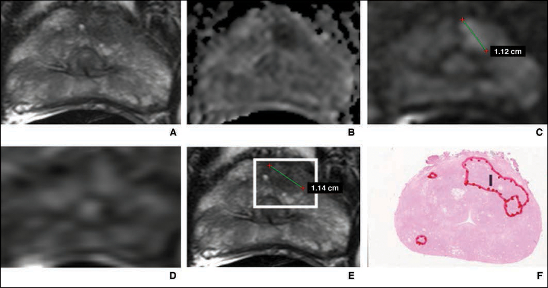
Fig. 1—

55-year-old man with prostate-specific antigen level of 4.68 ng/mL and Prostate Imaging Reporting and Data System category 5 lesion in left anterior transition zone correctly detected by artificial intelligence system. Final histopathologic result was Gleason 3 + 4 prostate cancer.
A, T2-weighted MR image.
B, Apparent diffusion coefficient map.
C, DW image (b = 2000 mm/s2).
D, Dynamic contrast-enhanced MR image.
E, T2-weighted MR image with attention box produced by means of artificial intelligence.
F, Photomicrograph of radical prostatectomy specimen. I = index lesion.
