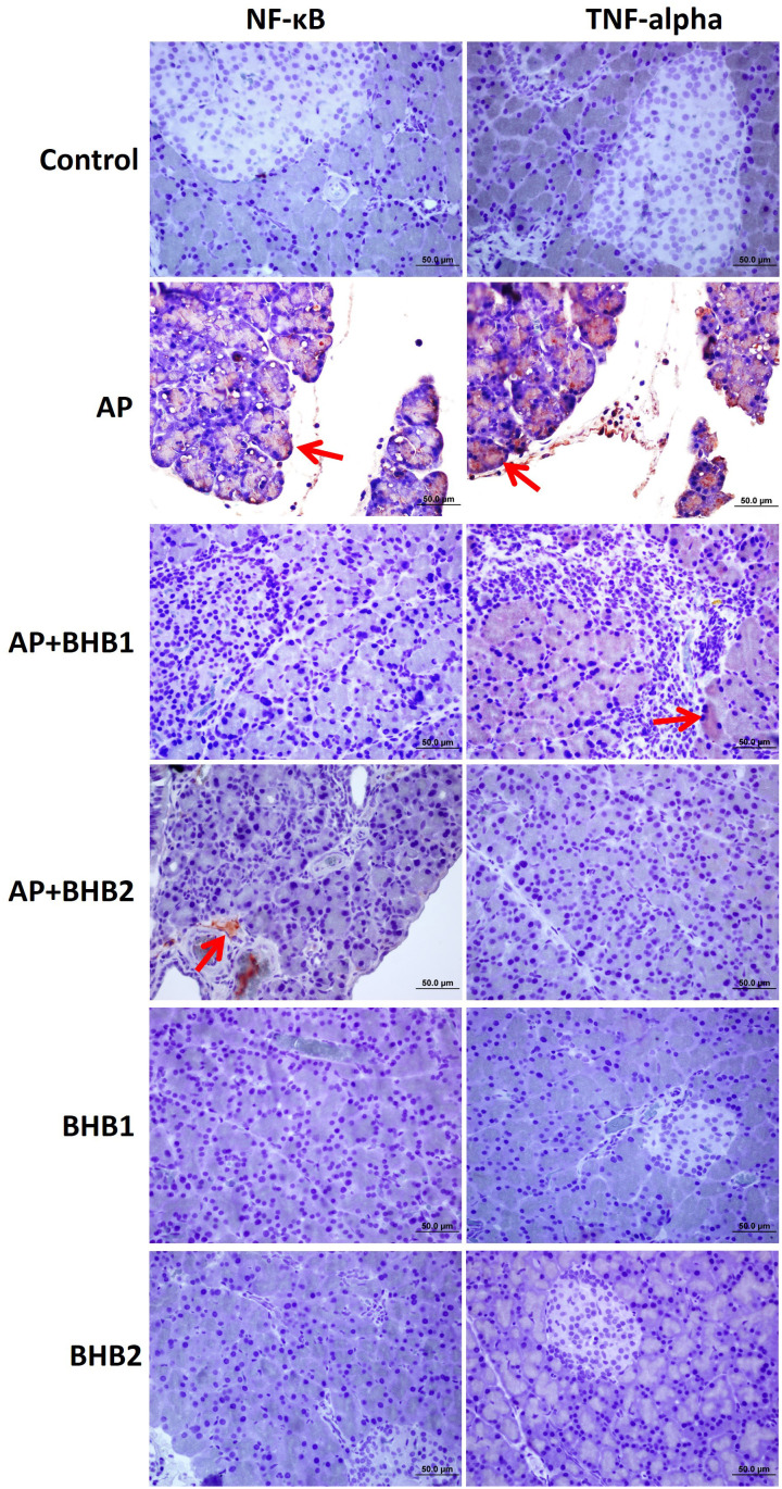
Figure 4.
NF-KB and TNF-α staining of pancreas sections. The BHB1 and BHB2 groups were poorly stained while the control group showed negative reaction for all antibodies. The most intense staining (red arrow) for all antibodies was detected in the AP group. It was determined that the immunoreaction for all antibodies was decreased in the AP + BHB1 and AP + BHB2 groups, when compared to the AP group.

