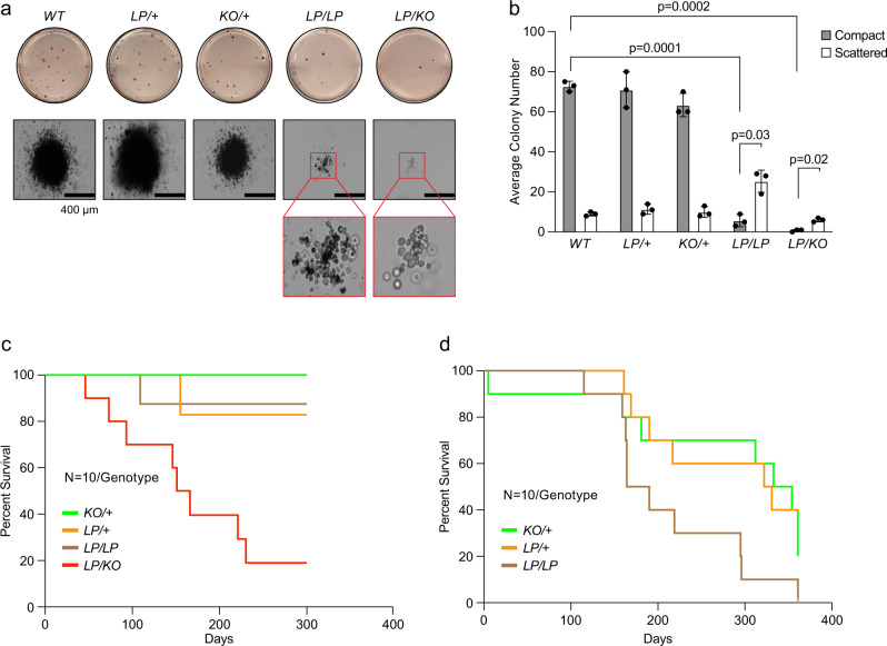
Fig. 2. Functional characterization of hematopoietic progenitor cells from fetal liver.
a Colony-forming assay to examine proliferative potential of hematopoietic progenitors from fetal liver isolated from 16.5dpc embryos of different genotypes. LP/LP and LP/KO showed reduced number of compact colonies. Inset shows morphology of scattered colonies. b Quantification of colony numbers for indicated genotype shows reduced number of compact LP/LP and LP/KO colonies and higher number of scattered LP/LP and LP/KO colonies (n = 3 biological replicate, two-tailed Students t-Test, error bar- SD of mean). c Kaplan–Meier survival curve of lethally radiated recipient mice after bone marrow reconstitution by fetal liver cells of indicated genotypes. LP/KO fetal liver cells show reduced bone marrow reconstitution leading to shorter median survival of the recipients (n = 10, p = 0.0003, Log-rank Mantel–Cox test). All other genotypes showed no significant difference in survival. d Kaplan–Meier survival curve of lethally radiated recipient mice after secondary transplantation using bone marrow from primary transplantation recipients. LP/LP bone marrow reconstitution shows significantly reduced median survival as compared to WT (n = 10, p = 0.041, Log-rank Mantel-Cox test).

