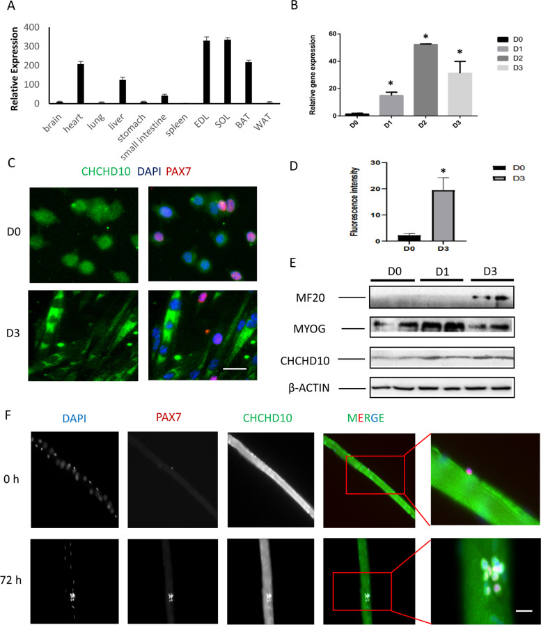
Fig. 1.
Chchd10 expression is enriched in skeletal muscles and upregulated during differentiation of satellite cell-derived primary myoblasts. A Relative mRNA levels of Chchd10 in various mouse tissue determined by qPCR, normalized to the mRNA level of the brain. N = 3. B Relative levels of Chchd10 mRNA in primary myoblasts at various days (D0-D3) after induced to differentiate. * p < 0.05 compared to D0 value. N = 3. C Immune staining of CHCHD10 (green) and PAX7 (red) in myoblasts before (D0) and after (D3) differentiation, scale bar: 20 μm. D Fluorescence intensity of CHCHD10 in myoblasts before (D0) and after (D3) differentiation. E Protein level of MF20, MyoG, CHCHD10 and β-ACTIN in D0, D1 and D3 differentiating myoblasts. F Immune staining of CHCHD10 (green) and PAX7 (red) in quiescent (0 h) and activated (72 h) satellite cells cultured on myofibers, scale bar: 20 μm. Nuclei were labeled with DAPI. The cluster of cells at 72 h contains self-renewed, proliferating and differentiated cells

