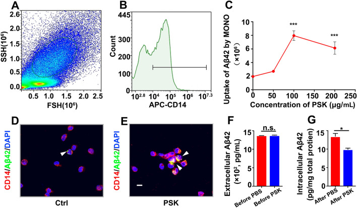
Fig. 1.
PSK enhances uptake and degradation of Aβ42 by monocytes in vitro. A, B Monocytes are selected by high-intensity CD14 labelling followed by gating of FSC-H and SSC-H. C Aβ42 uptake by monocytes versus dose of PSK. D, E Confocal stack images of Aβ42 uptake by human monocytes stained with Alexa594-conjugated anti-CD14 monoclonal antibody (red) and counter stained with DAPI (blue); FITC-conjugated Aβ42 is shown in green (scale bar, 10 μm). F, G Extracellular and intracellular Aβ42 levels before and after intervention assessed by ELISA (mean ± SEM. of triplicate wells in each treatment group). *P <0.05, ***P <0.001, n.s, not significantly different, one-way ANOVA and Student’s t-test. FSC, forward scatter; SSC, sideward scatter; PSK, polysaccharide kestin; Ctrl, control.

