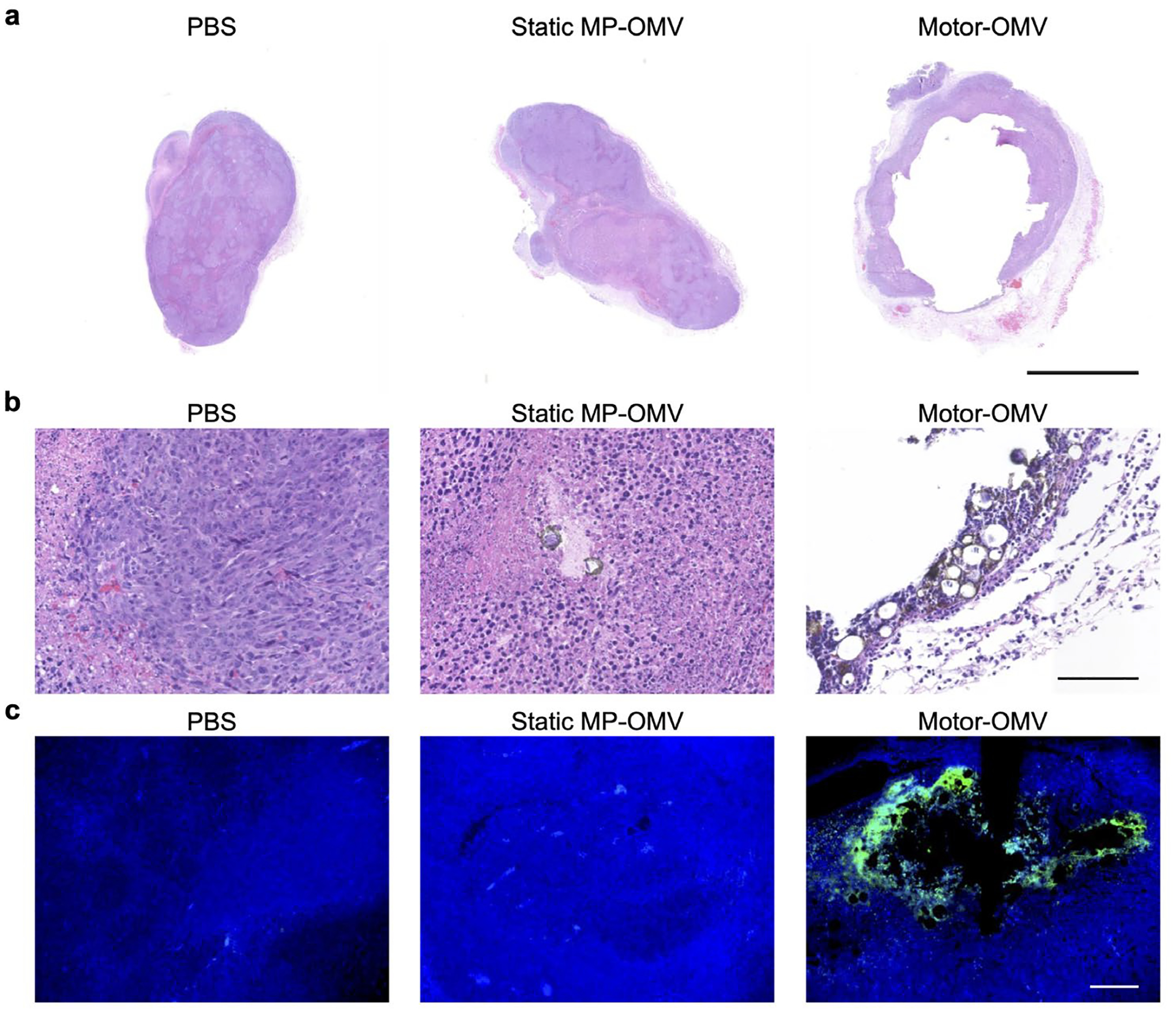
Figure 3.

Mechanical destruction of solid tumor tissue in vivo by Motor-OMV. a) Representative H&E-stained whole tumor sections taken 1 day after treatment with Motor-OMV or control samples (scale bar = 2.5 mm). b) Representative H&E-stained tumor sections at higher magnification with micromotors visible in brown (scale bar = 100 μm). c) Fluorescence visualization of representative tumor sections stained with TUNEL (green) and DAPI (blue) (scale bar = 100 μm).
