Figure 1.
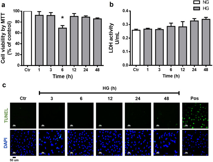
High glucose concentrations do not affect cell viability in MC. Cells were incubated with NG (Ctr; 5 mM glucose) or HG (25 mM glucose) for different time periods (1–48 h). (a) Cell viability was determined by the MTT assay; viability was expressed as the percentage of optical density respect to cells exposed to NG (100%). Cell death was measured through LDH release and TUNEL assay. (b) The results of LDH release are expressed as units per milliliter (U/ml). (c) Representative fluorescent micrographs of TUNEL-positive cells (green) compared to total cells (DAPI, blue) in MC exposed to NG or HG; positive control (pos: plus DNAse). Data are expressed as the mean ± SEM of duplicate cultures and are representative of three independent experiments. Scale bars: 50 µm. *p < 0.05 with respect to NG.

