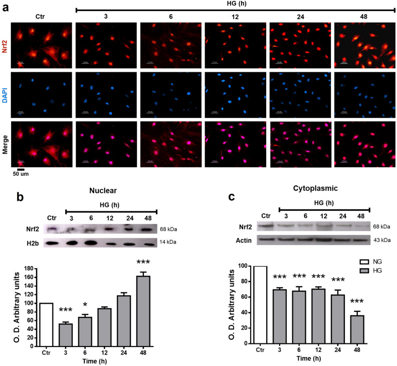
Figure 4.
Nrf2 subcellular localization in MC. Cells were exposed to HG for the indicated time intervals. (a) Immunofluorescent localization of Nrf2 in MC. Blue marks nuclei (DAPI); Red, Nrf2-staining, and Pink, merge of blue and red indicating nuclear localization of Nrf2. (b) Nuclear and (c) Cytoplasmic Nrf2 expression in MC exposed to HG. The upper part, representative western blot of Nrf2. The lower part, quantification of the relative levels of Nrf2. The relative expression levels were normalized using actin (cytoplasmic) or H2b (histone 2b; nuclear). Values represent the mean ± SEM (n = 5 per group) carried out in duplicate. NG, normal glucose; HG, high glucose. Scale bar represents 50 µm. *p < 0.05 with respect to NG; ***p < 0.001 with respect to NG. Full-size blots are presented in Supplementary Fig. 4.

