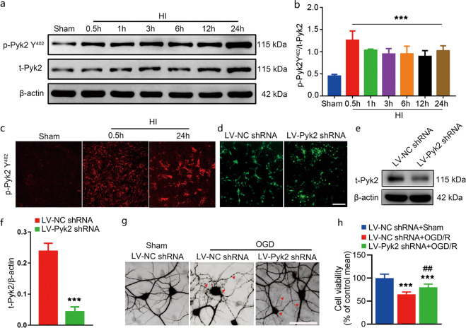
Fig. 1. Activation of Pyk2 after neonatal HI.
a The phosphorylation of Pyk2 at Y402 was measured by Western blotting 0.5, 1, 3, 6, 12, and 24 h after HI brain injury. b The time-course of changes in the ratio of phosphorylated Pyk2 Y402 and Pyk2 Y402. The data shown are means ± SD, n = 5 for each time point. ***P < 0.001 vs. the sham group. c Immunofluorescence staining of p-Pyk2 Y402 (red) in the cortex after HI. d Lentivirus infection of primary mouse cortical neurons was assessed by detecting GFP. e, f The inhibition of Pyk2 in neurons treated with viral vectors was evaluated by Western blotting. Pyk2 shRNA reduced total Pyk2 expression in Pyk2 shRNA-treated neurons. g The neuronal-specific marker MAP2 was used to identify primary mouse cortical neurons and assess their cytoskeleton morphology after OGD (bar = 5 μm). h Cell viability was assessed by the MTT assay after OGD/R. The data are presented as the means ± SD, n = 5. ***P < 0.001 vs. the LV-NC shRNA group in f and the LV-NC shRNA + sham group in h; ##P < 0.01 vs. the LV-NC shRNA + OGD/R group.

