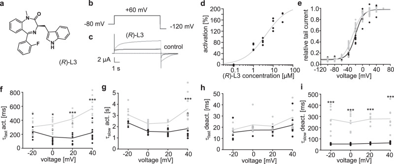
Fig. 1. (R)-L3 activates and slows the rates of activation and deactivation of hKv7.1 channels expressed in Xenopus oocytes.
a Chemical structure of (R)-L3. b Pulse sequences for voltage-clamp experiments. c Effect of 1 µM (R)-L3 on hKv7.1 currents, recorded in an oocyte by a 7-s pulse to potentials of −100 mV to +60 mV from a holding potential of −80 mV. Currents were recorded in control solution containing 0.1% DMSO followed by perfusion with 1 μM (R)-L3 containing solution. d Dose–response curve for (R)-L3 from hKv7.1 expressing oocytes at +40 mV test voltage. Each concentration was applied to four independent oocytes (n = 20) e Voltage dependence of current activation in the absence (black; n = 13) and presence of (R)-L3 (gray; n = 15) determined from peak tail currents measured at −120 mV. Currents were normalized to the peak tail currents elicited after a pulse to +40 mV. f–i Kinetics were evaluated in both absence (black; ctrl) and presence (gray; +(R)-L3) of 1 µM (R)-L3 and fitted by a two-component exponential function. Time constants are calculated for individual oocytes and given for fast (f; n = 9 for both conditions) and for slow (g; n = 9 for ctrl, n = 8 for + (R)-L3) component of Kv7.1 activation as well as for fast (h; n = 8 for ctrl, n = 6 for + (R)-L3) and for slow (i; n = 7 for ctrl, n = 8 for + (R)-L3)) component of Kv7.1 deactivation. Significance of mean differences was analyzed by one-way ANOVA and posthoc mean comparison Tukey test (*p < 0.05, ***p < 0.001).

