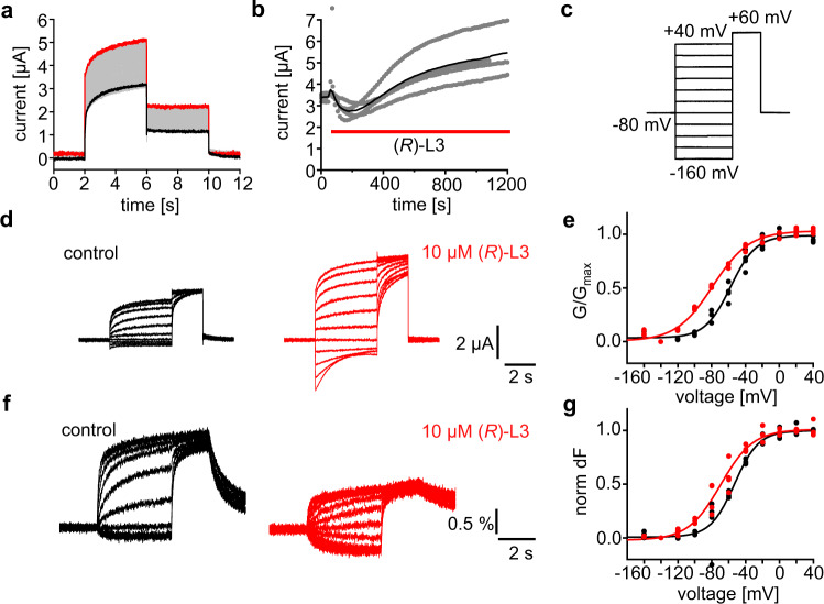
Fig. 3. (R)-L3 potentiates Kv7.1VCF (C214A/G219C/C331A) currents and left shifts the voltage-dependence of G/Gmax and dF/F.
a Whole-cell currents from an oocyte expressing Kv7.1VCF labeled with Alexa 488 C5 maleimide. Every 15 s, the membrane voltage was pulsed from the −80 mV resting potential to +60 mV for 4 s, followed by 2 s tails at −40 mV. Currents before (black) and after (red) a bolus of (R)-L3 was added to the bath (final concentration ~10 μM). b Steady-state current at +60 mV versus time during application of (R)-L3 (indicated by red bar). Blackline represents the mean value, gray dots represent raw data from each measurement (n = 4). c Pulse protocol for simultaneous current and fluorescence recordings in (d-g). d Sample current trace from a single oocyte before (black) and after (red) exposure to ~10 μM (R)-L3. e G/Gmax voltage relationship of Kv7.1VCF expressing oocytes in the absence (black) and presence (red) of (R)-L3 (n = 4 for both conditions). f Sample fluorescence trace from an oocyte before (black) and after (red) exposure to ~10 μM (R)-L3. g dF/F voltage relationship of Kv7.1VCF expressing oocytes in the absence (black) and presence (red) of (R)-L3 (n = 4 for both conditions).

