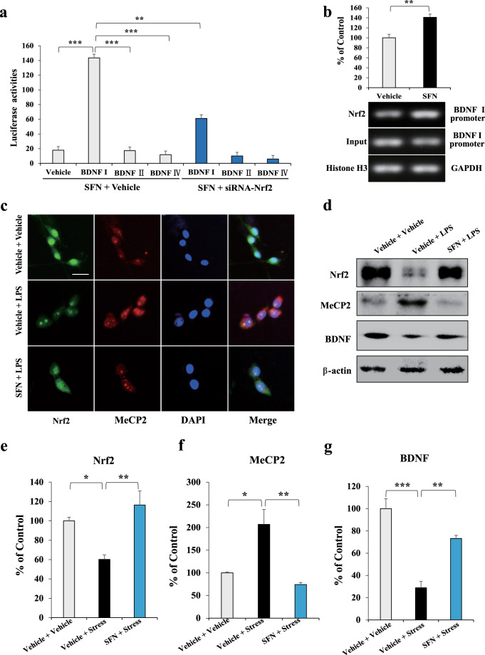
Fig. 3. Activation of Nrf2 by SFN results in BDNF transcription in BV2 cell.
a The luciferase reporter assay for the activation of BDNF I, II, and IV promoter. BV2 cells are treated with SFN or siRNA-Nrf2 for 24 h followed by luciferase examination. Data of the pcDNA, BDNF II, and IV promoters are compared with those of BDNF I promoter (mean ± SEM, n = 8 per group, one-way ANOVA, **P < 0.01, ***P < 0.001). b ChIP assay for the BDNF I promoter. The Nrf2 protein–DNA cross-linking samples are obtained from BV2 cells treated with SFN or vehicle via co-immunoprecipitation with anti-Nrf2 antibody. PCR is carried out with the BDNF exon I promoter primers (mean ± SEM, n = 4 per group, Student’s t test, **P < 0.01). c Immunofluorescence staining for Nrf2 and MeCP2. BV2 cells are treated with SFN or LPS for 24 h followed by immunofluorescence staining. Scale bar, 50 μm. d Representative images of the Western blot analysis for Nrf2, MeCP2, and BDNF. Quantifications of Nrf2 (e), MeCP2 (f), and BDNF (g) in Western blot analysis. (Mean ± SEM, n = 4 per group, one-way ANOVA, *P < 0.05, **P < 0.01, and ***P < 0.01).

