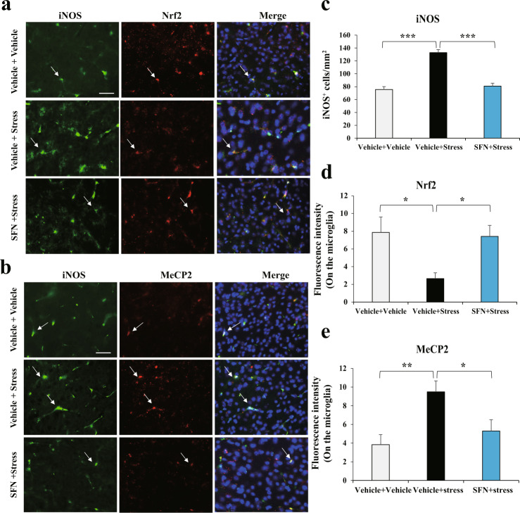
Fig. 5. SFN suppresses stress-induced upregulation of iNOS-positive microglia, which is associated with changed Nrf2 and MeCP2 abnormal expression in microglia with pro-inflammatory phenotype in stressed mice.
a SFN suppresses stress-induced upregulation of iNOS-positive microglia, which is associated with increased Nrf2 expression in iNOS-positive microglia. Representative images of iNOS and Nrf2 in the mPFC of stressed mice. Representative positive stainings are indicated by arrows. Scale bar, 50 μm. b SFN suppresses stress-induced upregulation of iNOS-positive microglia, which is associated with decreased MeCP2 expression in iNOS-positive microglia. Representative images of iNOS and MeCP2 in the mPFC of stressed mice. Representative positive stainings are indicated by arrows. Scale bar, 50 μm. Quantification of iNOS (c), Nrf2 (d), and MeCP2 (e) fluorescent intensity in the iNOS-positive microglia (Mean ± SEM, n = 4 per group, one-way ANOVA, *P < 0.05, **P < 0.01, and ***P < 0.001).

